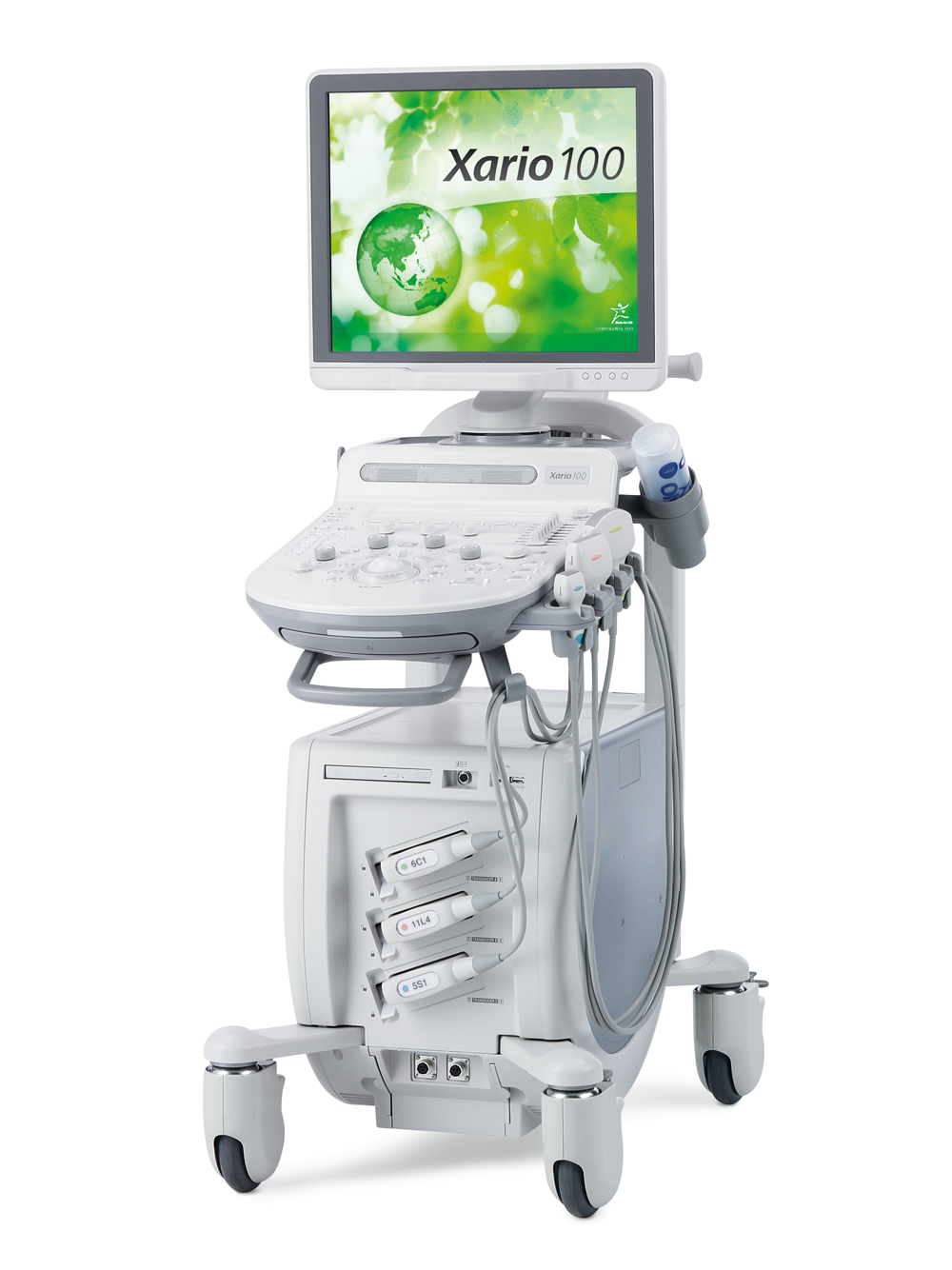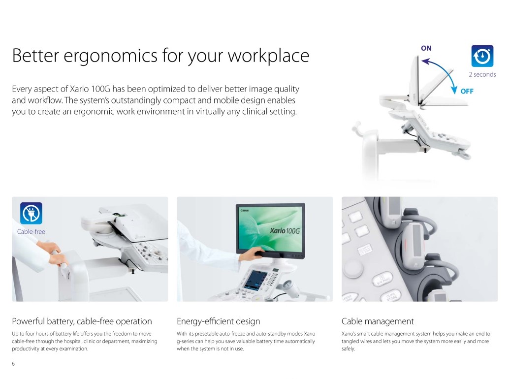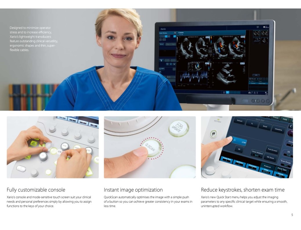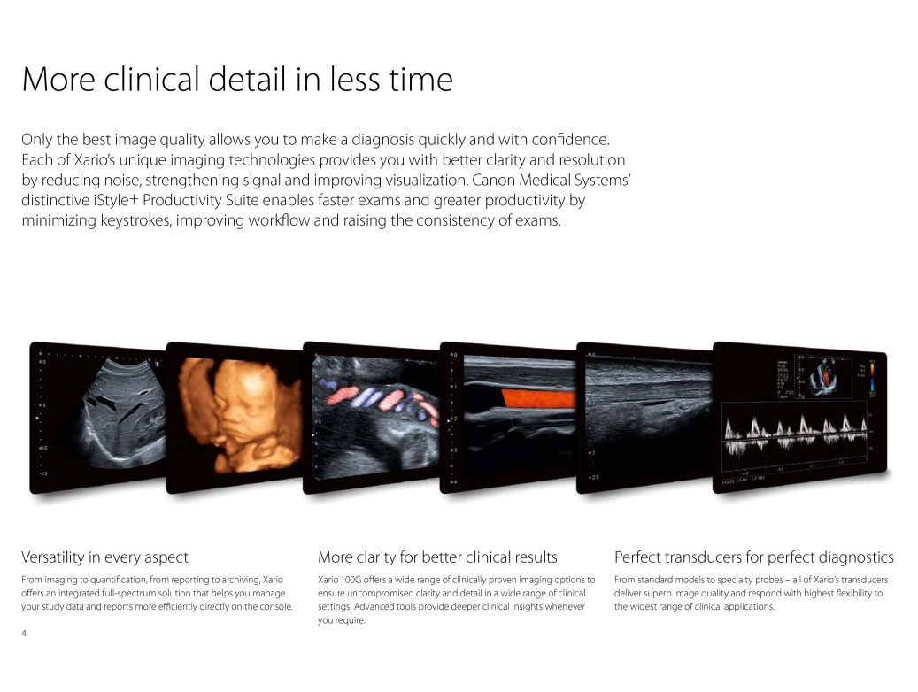
Xario 100 and 200’s High-Density Beamformer Architecture produces advanced digital signal processing technology to reveal clinical information never seen before. With comprehensive image enhancement capabilities and an unsurpassed 40 cm depth setting, Canon/Toshiba is clearly the leader in delivering best-in-class images for all patient exams.
clearly the leader in delivering best-in-class images for all patient exams.
Features/Specs
19” High Resolution IPS Monitor
Fully Customizable, Mode Sensitive Touch Screen
Three Transducer Sockets
Super High Density Transducers
Optimized Workflow
Comprehensive on-board reporting
DICOM® 3.0 Connectivity
Export Images Using USB, Network or Built-in DVD Drive
Advanced Features
Precision Imaging enhances the definition and sharpens the edges of structures to separate clinical information from noise.
Differential Tissue Harmonic Imaging (D-THI) increases contrast and spatial resolution at greater depths and on difficult-to-image patients.
ApliPure+ uses a new generation of compound imaging technology to achieve unparalleled uniformity and detail while preserving clinically significant markers.
Advanced Dynamic Flow™ (ADF) displays smallest blood vessels and complex blood flow with unequaled accuracy and detail.
Volume Imaging Suite captures volume data sets at high-volume rates for shorter exam times and features a comprehensive set of imaging modes (Surface rendering, Multiview and MPR).
Stress Echo enables fast and accurate wall motion assessment and supports both standard and user-definable protocols for exercise and pharmacological stress studies.
Real-time Elastography provides a visual representation (color mapping) of the elasticity of lesions following manual compression and helps localize and assess palpable masses with exceptional accuracy, sensitivity and reproducibility.
Panoramic View helps visualize widespread areas and anatomical relationships by creating wide-view images of a region of interest.
BEAM (Biopsy Enhancement Auto Mode) provides a clearer visualization of biopsy needles in the live ultrasound image. BEAM enhances the visibility of a biopsy needle and works with all common needle sizes. The function provides three enhancement levels and selects the best scan angle fully automatically.


